Transmission Electron Microscope – Working, Use Cases, Comparison with SEM
Out of all the instruments used for examining tiny details, few are as powerful as the Transmission Electron Microscope (TEM). It is capable of viewing things from viruses, through complex cell structures and to nanomaterials. Surpassing the limits of conventional light microscopes, TEM truly opens a whole new world.
In this article, we will discuss what TEM is and how it works, how it compares to SEM (Scanning Electron Microscope), and the appropriate times to utilize it. You will also gain insights into the instrument’s structure and principles, as well as what makes it one of the most essential instruments in science and engineering.
What is TEM and How Does it Work?
TEM, short for transmission electron microscope, is a high resolution imaging tool which uses electrons in place of light to visualize ultra-fine details of specimens.
Here’s the process:
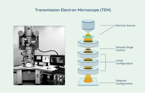
Step 1: Electron Source: Starts with a beam of high energy electrons
Step 2: Electron Beams: Focused by condenser lenses
Step 3: Specimen Interaction: Pass through an ultra thin specimen layer. Some electrons are scattered while others are transmitted.
Step 4: Image Formation: A magnified image is formed by hitting the fluorescent screen or camera sensor with transmitted electrons.
Using electrons instead of light allows construction of structures as small as 0.1 nanometers. This makes it possible for scientists to view structures and details at an atomic level.
What is the basic principle of a TEM?
Electrons are the basis of a TEM. They go through a specimen and create images based on the core principles below:
- Electrons have wave properties and can be concentrated using lenses.
- Region within the specimen is capable of being permeated by electron beams, permitting internal electron-driven interactions
- The scattering of electrons from a sample structure causes the image to appear differently.
TEM makes it possible to see very detailed images of the internal structure of cells, materials, and molecules.

What is the structure and function of TEM?
The construction of a transmission electron microscope involves several parts. The function of the highlighted pieces is below:
1. Electron Gun
An electron beam is produced from a tungsten filament cathode because of high current, or from a field emission source.
2. Condenser Lens System
Focuses and adjusts intensity to the electron beam placed on the specimen.
3. Specimen Holder
Holds the specimen which thickness is in the order of 100 nanometers.
4. Objective Lens
Collects electrons from the sample and forms the first image. This is done by the lens.
5. Intermediate and Projector Lenses
Further increase the size of the image and project it onto a screen or into a camera.
6. Viewing Screen / CCD Camera
Presents or captures the last image, so that it can be digitised and analysed scientifically.
All parts together to assist TEM in visualizing the intricate internal elements of a component at an atomic level.
What are the things viewed with a TEM?
TEM enables the visualization of very small features within materials and biological samples, such as:
- Cell organelles like mitochondria
- Viruses and bacteria
- Defects on crystal lattice in metals and ceramics
- Nanoparticles
- Proteins and organized molecules
TEM can achieve magnification of 2 million times, revealing fine features that are utterly invisible with a standard microscope.
When would you need to use a TEM microscope?
In fields requiring high precision and internal imaging, TEM is preferred. A few practical applications are:
- Cell biology – study structures of cell organelles
- Virology – observe viruses in detail
- Material science – find grain boundaries, dislocations and defects
- Nanotechnology – investigate nanoparticles and their activity
- Forensics – trace material residue
When examining a thin specimen’s internal ultrastructure at an atomic or subatomic level, TEM is the most suitable instrument.
Which aspects differentiate TEM from SEM?
TEM and SEM are two types of electron microscopes but they operate differently, so they have different applications. Here is a overview:
| Feature | TEM | SEM |
| Image Formation | By electrons transmitted through the sample | By electrons scattered off the surface |
| Sample Requirement | Must be very thin | Can be bulkier or thicker |
| Resolution | Higher (up to 0.1 nm) | Lower (1–20 nm) |
| Image Type | 2D internal structure | 3D surface topography |
| Usage | Biology, materials, nanotech | Surface morphology, coatings, biology |
When would ultimately TEM be more useful than SEM?
In comparison to SEM, TEM would be more useful when you need to view:
- Substructures within a cell or piece of material
- The crystalline structure, and possibly its lattice defects.
- Viruses and macromolecules to a very capable resolution (extremely detailed).
- Extremely fine parts.
In comparison, SEM works best with examining outer surface features while TEM works best with inner features.
What are the main advantages of TEM over SEM?
- Unmatched resolution which enables imaging of features to atomic levels.
- Internal imaging shows structures inside whereas SEM cannot.
- Material characterization techniques
- Extremely precise description of the crystal’s structure by means of electron diffraction.
- Able to image required parts of a specimen further than SEM by up to 2 million times.
These benefits offer advantages in advanced research and contexts requiring utmost precision are of particular importance.
What is a transmission electron microscope quizlet?
A Transmission Electron Microscope Quizlet refers to any flashcard or study set that was specifically made on the Quizlet platform. These usually contain:
- Identification of the parts of the TEM
- Function of each part
- Differences of TEM to SEM
- Use or relevance in real life
Through these, learners increase their understanding of the different concepts in microscopy and bolster their preparedness for the relevant assessments.
Challenges and limitations of TEM
Even though TEM offers powerful capabilities, it has certain challenges. These include:
- Highly expensive equipment along with frequent maintenance.
- Complicated structural metal sample preparation.
- Fewer than 100 nanometers thickness sample size.
- Not applicable for living things.
- No untrained personnel TM.
- No untrained full TM.
With regard to these challenges, the benefits of TEM are far greater than the challenges in research and discovery worthy settings.
Real world application of TEM
TEM has impacted many fields of science:
Medicine and Biology
- Virus structure discovery of viruses such as covid 19.
- Cell mutation and disease progression study.
Material sciences
- Stress fractures and alloy composite metals.
- Stronger lighter alloy composites.
Chemistry
- Nanoparticle structure analysis.
- Catalyst behaviour under atomic observation.
Semiconductor Industry
- Transistor channel and integrated circuit imaging.
Due to its unparalleled ability to provide nanometer and even sub-nanometer level resolution, TEM is critical in advancing modern technology.
The contributions of transmission electron microscope to contemporary science
Some of its contributions are well known. Without TEM, the detailed structure of the following would remain a mystery:
Molecular structures of DNA and proteins
- Viral capsids and envelopes
- Crystalline structures of metals
- Carbon nanotubes and graphene
Its achievements in pharmaceutical and drug research, along with vaccine development, and even in space technology materials, make it an unsung champion of scientific endeavors.
How do you decide whether to use a Transmission Electron Microscope for your research?
If you need to study viral structures, look into physical defects of materials, or analyze nano-engineered components, no other tool can provide the clarity and resolution that TEM offers.
It is due to this unparalleled capacity to depict the undepictable that formed the basis for disciplines, spanning biomedicine to aeronautics. Not every sample may be appropriate for TEM, but wherever precision is critical, comfort is found.
If there ever arose a difficult question “What does a TEM microscope let you see?”, the answer would be almost everything that cannot be reached by human sight, and at times, even more than that.


Post Comment